
 Aqueous Humor - The fluid produced within the front part of the eye.
Bleb - A bubble within the tissue overlaying the new drainage opening created during surgery.
Central Vision - What is seen when you are looking straight ahead or reading.
Cilliary Tissues - Tissues located around the lens of the eye that supply nourishing fluid to the eye.
Cones Light - sensitive retinal receptor that provides sharp visual acuity and color discrimination, most numerous in macula area.
Conjunctiva - A thin, clear membrane that lines the inner surface of the eyelids and the outer surface of the eyeball, except for the cornea.
Cornea - The clear part of the eye located in front of the iris. Part of the eye's protective covering.
Crystalline Lens - The eye's natural lens. Transparent, biconvex intraocular tissue that helps bring rays of light to focus on the retina.
Ciliary Muscles - Smooth muscle portion of the ciliary body. Responsible for relaxation of zonules, to allow the crystalline lens to focus.
Drainage Canals - Small openings around the outer edge of the iris, which provide the final pathway for fluid to leave the inside of the eye. Sometimes referred to as the trabecular meshwork or, Schlemm's canal.
Filtering Bleb 5-FU - A medication that stops the healing process. Sometimes used around the new drainage hole created during surgery (the bleb) to stop it from healing or scarring over.
Fovea - pit in the macula that produces sharpest vision.
Glaucoma Suspect - A person may be considered a glaucoma suspect on the basis of high intraocular pressure, a suspicious appearance of the optic disc or visual field, a family history of glaucoma, or narrow angles between the iris and cornea.
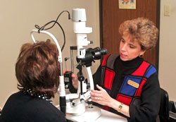
Gonioscopy - In this test, a contact lens that contains a mirror is gently placed on the eye. The mirror lets the doctor look sideways into the eye to check whether the angle where the iris meets the cornea is open or closed. This helps the doctor decide whether open-angle or angle-closure glaucoma is present.
 Intraocular Pressure (lOP) - The internal pressure of the eye. Normal intraocular pressure usually ranges from 10-22 mm Hg, although people with relatively low pressures can still have glaucoma.
Iris - The pigmented tissue lying behind the cornea that gives color to the eye. The iris can expand or contract to allow just the right amount of light to enter the eye.
Lens - Located behind the iris, helps light focus onto the retina.
Macula Small - central area of the retina, area of acute central vision. Used for reading and discriminating fine detail and color.
Microsurgery - Surgery performed with a microscope in which an instrument is used to make a tiny, new opening in the sclera so that intraocular fluid can drain out of the inside of the eye.
Mm Hg - An abbreviation for "millimeters of mercury," which is a scale for recording intraocular pressure.
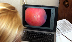
Ophthalmoscopy - A test designed to examine the inside of the eye, especially the optic nerve. An instrument with a small light on the end is held up to the inside of the eye in a darkened room. This instrument lights up and magnifies the eye, and lets the doctor look at the shape and color of the optic nerve.
Optic Nerve - Second cranial nerve. Largest sensory nerve of the eye, carries impulses for sight from the retina to the brain. It is the nerve in the back of the eye that carries visual images to the brain.
Perimetry - Also known as the visual field test. A test that produces a map of the complete field of vision, designed to determine whether there is damage to any area of vision.
Peripheral Vision - The top, sides, and bottom areas of vision, which are usually the first areas of vision affected by glaucoma.
Pupil - Variable sized black circular opening in the center of the iris that regulates how much light enters the inner part of the eye.
Retina - The part of the eye that carries light and images to the brain through the optic nerve.
Sclera - The tough, white, protective, outer coat of the eye.
 Tonometry - The use of a device to measure the pressure within the eye.
Trabecular Meshwork - The formal name of the mesh-like drainage canals all around the iris.
Vitreous Body - Chamber that is located behind the lens and in fornt of the retina, contains clear, gelationousfluid called vitreous humor.
